


Harnessing Graphene for Precise Radiotherapy Skin Dosimetry in Nasopharyngeal Cancer

Introduction
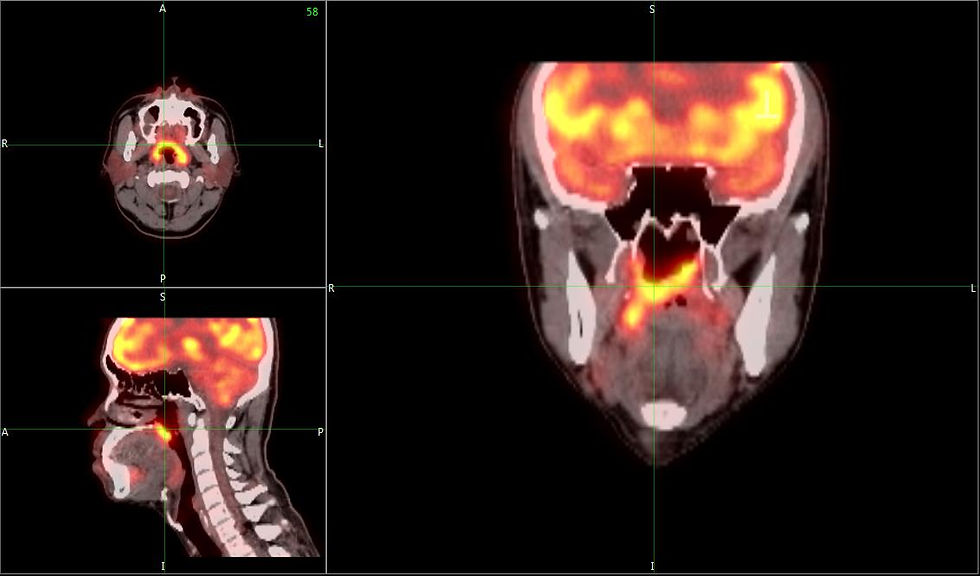
Nasopharyngeal carcinoma (NPC) is the cancer of the nasopharynx, constituting the passageway between the nose and the back of the mouth.
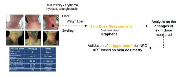
Although accounting for a lower global incidence of NPC (1 in 100,000), it is statistically prevalent in the Southeast Asian, Middle Eastern, and South African regions, with approximately 15-30 cases per 100,000. Radiotherapy remained the mainstay treatment for early or locally advanced NPC due to the proximity of the nasopharynx to delicate structures within the neck, thereby limiting its surgical access. NPC treatment typically requires 35 consecutive days of radiation therapy. However, significant weight loss and skin toxicity effects arising from radiotherapy among patients during treatment can alter their anatomy, impacting the precision of treatment planning and potentially compromising the effectiveness of radiation delivery.
Gathering a team of remarkable scientists from the Department of Physics, the Department of Oncology, and the Department of Biomedical Imaging, the research team led by Associate Professor Dr. Siti Fairus from the Department of Physics at the Universiti Malaya (UM) successfully developed graphene-based materials capable of precise radiotherapy dose calculations for NPC.
Setbacks in the Current Provision of Radiotherapy
As previously introduced, dosage precision in current radiotherapy treatments for NPC is inevitably impacted by anatomical changes resulting from its side effects. Hence, Adaptive Radiotherapy (ART) was introduced in the current course of radiotherapy to tailor the treatment to a patient’s anatomical changes over time. However, as shared by Associate Prof Dr. Siti Fairus, several limitations impact the accuracy of dose calculations achieved under ART:
● Imaging Challenges: High-quality imaging is crucial for accurately delineating tumour and organ-at-risk (OAR) structures. While modalities like Cone Beam Computed Tomography (CBCT) are commonly used, they may suffer from lower image quality than traditional CT scans, potentially compromising the precision of dose calculations.
● Dose Accumulation Complexities: Accurately summing doses from different treatment sessions is essential to ensure optimal delivery and adherence to dose constraints. However, due to the complexities of mapping anatomical changes, inaccuracies in deformable image registration (DIR) can lead to errors in dose accumulation.
● Resource Constraints: Implementing ART requires significant resources, including advanced imaging technologies and trained personnel. A multi-centre survey in Malaysia revealed that while many centres recognise the benefits of ART, limitations such as equipment availability and workflow integration hinder its widespread adoption.
Development of Graphene-Based Materials
The journey toward developing graphene-based dosimetric tools was first ignited by exploring the component of pencil lead - Graphite1. With a carbon content of 98%, graphite closely mimics the elements found in human skin, which also have a high carbon composition. This similarity provided a foundation for its use in dosimetry.
An earlier study, conducted in collaboration with Sunway University, explored the potential of graphite for radiation dose measurement and reported promising results, despite the limitations of the layered structure of graphite in terms of sensitivity and adaptability. This success inspired a shift to graphene, a single atomic layer of carbon derived from graphite. Graphene’s structure provides superior properties, including higher sensitivity, increased conductivity, and a more robust ability to detect radiation changes in real-time. These advancements made graphene a natural evolution of the dosimetric material.
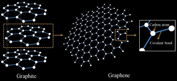
The value of graphene as a dosimetric material is further amplified by its biochemical compatibility. This is evidenced by the high carbon content and an effective atomic number (Zeff = 7.4), which is equivalent to that of human soft tissue and closely mimics the interaction of radiation with human tissues. This property contributes to the safety and effectiveness of graphene upon clinical application.
Another driving force behind this research is the critical need to address the side effects of radiation therapy for nasopharyngeal carcinoma (NPC) patients. NPC treatments typically require 35 consecutive days of radiation therapy, delivering a total dose of 70 Gy. However, significant weight loss among patients during treatment can alter their anatomy, impacting the precision of treatment planning and potentially compromising the effectiveness of radiation delivery.
By developing graphene-based dosimetry tools, this project aims to provide real-time adaptive monitoring, continuously optimising the radiation dose and thereby reducing potential side effects and improving treatment outcomes for NPC patients.
1Graphite - A stack of tightly bound carbon sheets
2Graphene - Derived from graphite; single atomic layer of carbon
How it Works
This project utilises graphene nanoplatelets as dosimeters for measuring skin dose during radiotherapy. Data obtained via nanoplatelets enables precise radiation monitoring delivered to the skin, allowing the identification of a trigger point attributed to altered anatomy, patient weight loss, or tumour shrinkage. As such, the identification and analysis of trigger points enable real-time dose validations and adjustments required in the provision of radiotherapy, leading to a reduction of unwanted side effects.
Key Discoveries & Challenges
The development of graphene-based dosimetry tools was accomplished through synergistic efforts from the Department of Oncology, which has expertise in analysing anatomical changes in NPC patients; the Department of Physics, which developed a graphene-based dosimeter; and the Department of Biomedical Imaging, which curated a predictive model for ART adjustments with the integration of artificial intelligence.
Nevertheless, every pursuit comes with its own set of growth challenges. The primary challenge faced by the research team was ensuring the consistent fabrication of graphene nanoplatelets. This was overcome with intensive training to produce uniform and high-quality graphene suitable for dosimetry. Additionally, to maintain the homogeneity of graphene films, the team refined the deposition techniques and controls of process parameters to achieve layers of consistent quality, ensuring measurement accuracy.
Through consistent refinement and perseverance, graphene-based dosimetry has been found to offer a cost-effective alternative to traditional methods, reducing equipment and operational costs while improving treatment outcomes. Its ability to monitor skin dose instantaneously enhances tumour control and minimises side effects, significantly improving the prognosis for NPC patients. Beyond radiotherapy, graphene's potential extends to biosensors, drug delivery systems, tissue engineering, and wearable health devices, thus promising transformative advancements in clinical practice.
Conclusion
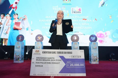
Ultimately, developing a real-time, carbon-based dosimeter addresses a critical gap in radiation therapy in NPC patients, enabling accurate skin dose measurements. Identifying trigger points for ART introduces an opportunity for personalised medicine, enabling effective patient care, as dose adjustments resulting from graphene-based dosimetry differ from person to person. The integration of AI in enhancing predictive capabilities further inspires innovation and revolutionary progress within the field of medicine. This novel and bold endeavour by Associate Prof Dr. Siti Fairus was recognised as the recipient of the prestigious “Anugerah Saintis Muda Negara 2024” by the Ministry of Science, Technology, and Innovation (MOSTI) in the Physics category. Through rigorous testing and approaches undertaken, the research confirms the potential of graphene nanoplatelets as a versatile material in a wide range of medical applications, offering a unified care approach that is adaptable, precise, and cost-effective for future healthcare.
Researcher featured:
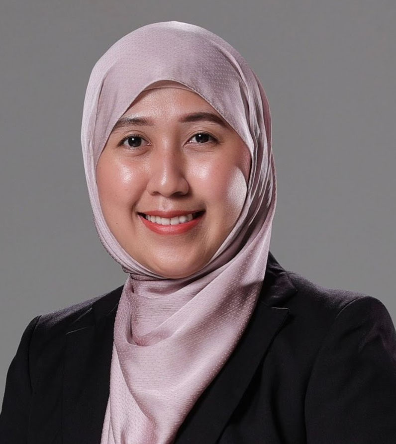
Assoc. Prof. Dr. Siti Fairus Abdul Sani
Department of Physics
Faculty of Science, UM
For inquiries, please contact:
T: +603-79674285
Author:

Ms Eng Pink Huey
Pink Huey is currently a fourth-year medical student at the Universiti Malaya. Outside clinical wards, her enthusiasm for writing and classical music drives her appreciation for the harmonious union of art and life. For a tinge of adrenaline, she enjoys hiking and chasing sunsets!
Copyedit:
Siti Farhana Bajunid Shakeeb Arsalaan Bajunid, Assistant Registrar, UM
Nurhazrin Zanzabir, Assistant Administrative Officer, UM


Comments